|
|
|
|
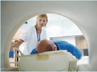
High field large bore 3T MR system |

|

|
|
|

Questions et réponses (FAQ)
Madame, Monsieur, vous allez bientôt subir un examen d'Imagerie par Résonance Magnétique (IRM), une technique irremplaçable. Sans danger pour l'organisme, des images très précises de votre corps vont être obtenues et analysées pour permettre à votre médecin traitant de mieux déterminer l'origine de vos troubles.
L'appareil illustré ci-dessus dispose du plus haut champ magnétique couramment disponible (3 Tesla), mais bénéficie d'une large ouverture agréable, limitant la sensation de confinement. Le bruit est réduit et la durée d'examen nettement raccourcie. Les images sont d'une très grande précision pour tout type d'examen.
Avant l'examen:
- Vous n'avez pas besoin, sauf exception, d'être à jeun et vous pouvez prendre vos médicaments habituels.
- Certains fards contenant des métaux, il est recommandé de ne pas se maquiller avant un examen de la tête. Au besoin, vous pouvez nous demander un démaquillant.
- En arrivant, on vous demandera de vous déshabiller (le soutien-gorge aussi, car il contient des attaches métalliques) et de revêtir une blouse. Tous les objets métalliques, y compris les lunettes, la montre, les cartes de crédit (risque d'effacement!) seront déposés dans un casier fermant à clé. De même pour les appareils dentaires amovibles et les appareils d'aide à l'audition.
- Si vous le souhaitez, une personne de votre entourage peut rester avec vous durant tout l'examen.
Précautions:
- Par principe, on évite d'examiner les femmes enceintes au premier trimestre de la grossesse sans nécessité absolue. En cas de doute, l'examen pourra sans problème être reporté.
- Les personnes porteuses d'un stimulateur cardiaque (pacemaker), d'un défibrillateur, d'une pompe à insuline ou d'un appareil acoustique doivent éviter de rentrer dans l'enceinte de la résonance magnétique.
- Nous vous prions de signaler avant l'examen tout corps métallique implanté à la suite d'une opération ou d'un accident, en particulier dans la tête et près des yeux.
- Certaines valves cardiaques peuvent être incompatibles dans les IRM de très haut champ, veuillez vous renseigner à l'avance sur le type de valve que vous portez.
Déroulement de l'examen:
Couché(e) sur la table d'examen, la partie de votre corps à examiner sera positionnée au centre d'une antenne de réception. Une immobilité complète vous sera demandée. Quelle que soit la durée de l'examen (en général de 20 à 30 minutes), vous ne serez jamais laissé(e) sans surveillance. Le bruit saccadé produit par l'appareil est normal, il est dû à l'inversion rapide du courant dans les bobines. Si ce bruit vous incommode, vous pouvez demander des tampons auriculaires.
Parfois, pour obtenir plus de renseignements, il est nécessaire d'injecter dans une veine un produit de contraste; cette substance (Gadolinium) ne contient pas d'iode et n'a pratiquement pas d'effets secondaires.
Voici, en quelques mots, le principe de l'IRM:
Le corps entier du patient est soumis à un champ magnétique, tel que celui du globe terrestre ou d'un aimant, mais beaucoup plus intense. L'eau du corps humain contient des atomes d'hydrogène (protons), qui s'alignent dans ce champ magnétique. Pour obtenir un signal, l'appareil doit modifier localement l'orientation de ces protons au moyen d'impulsions d'ondes radio. Les protons aimantés ont vite tendance à reprendre leur orientation initiale, tout en émettant à leur tour des ondes radio de fréquence différente. Ces ondes sont captées par une antenne, analysées et reconstruites sous forme d'images en coupes, et ceci dans n'importe quel plan de l'espace (contrairement aux coupes uniquement transverses du scanner). La résonance magnétique n'utilise aucune radiation ionisante (rayons X), et personne n'a pu démontrer à ce jour le moindre effet nocif sur l'organisme dans dans des conditions d'examen habituelles. Le champ magnétique interagit cependant avec les objets métalliques, ce qui peut dégrader les images. Tout ce qui contient du fer est fortement attiré par l'aimant, d'où les précautions mentionnées plus haut.
Frequent asked questions
Madam, Sir, you will soon have a Magnetic Resonance Imaging (MRI) examination, a highly efficient imaging technique. This harmless diagnostic tool will provide very precise images of your body. After interpretation of theses images by the radiologist, your attending physician will better understand the nature of your disorder.
The illustrated MRI is a high field 3 Tesla MRI with large opening fitting evry patient with a lot of space in front of the patient's face; feelings of claustrophobia are minimized, a more comfortable positioning is possible and patient access is much easier. Moreover, the machine is definitely less noisy and examination time is shortened.
Before the examination:
- Unless specified, you may eat and take your usual drugs before MRI examination.
- As most make-ups are containing metal components, it is recommended not to make up before head or neck MRI. A make-up remover will be provided if necessary.
- After registration, you will be asked to undress (included brassiere, which contains metal fasteners) and to wear a blouse. All metal objects, including eyeglasses, watch, credit cards (risk of erasure!) will be removed and kept in a safe place. Any dental device should be removed if possible, as well as hearing aids.
- A friend or a parent may stay with you during your examination if you like.
Precautions:
- As a rule, pregnant women should not be exposed to MRI during the first trimester of pregnancy without peremptory necessity. If you think you could be pregnant, it is safer to postpone your examination until next period.
- If you are wearing a pace-maker (heart stimulator), an implantable defibrillator, an insulin pump or a fixed acoustic device, you are not allowed to enter the MRI room.
- Please mention before examination any metal foreign body resulting from an operation or an accident, particularly in your head or in the vicinity of your eyes.
- Some cardiac valves may be limited in very high field MRIs; please require valve information from your surgeon if possible.
During the examination:
Installed in a comfortable recumbent position, the patient body part to be examined will be positioned in the center of a reception antenna. Complete immobility will be required during the sequences. Whatever the duration of the examination (usually from 30 to 50 minutes), you will never be left without supervision. The repetitive noise you hear is normal, it is due to fast current inversions in the gradient coils. If this noise bothers you, just ask for ear plugs.
To obtain additional information, a contrast agent must sometimes be injected into a vein; this substance (Gadolinium) is non toxic, does not contain iodine and side effects are exceptional.
MRI principles summary:
MRI (magnetic resonance imaging) is performed with examined part of the patient centered in a magnetic field identical but much stronger than the field generated by the earth or by a pocket magnet. Most hydrogen atoms (protons) contained in body water tend to fall into line with this magnetic field. Pulsed radio waves of short duration are then locally applied to modify proton orientation. Protons tend to return to their initial alignment, with emission of radio waves of specific frequency; this emission is detected by an antenna placed around the patient and computed into images; MRI views may be obtained in any plane of space, whereas CT-scan native images are only transverse. MRI does not generate any X-rays and no harmful biological effects have ever been proved under standard examination conditions. However, magnetic field interacts with metallic objects, resulting in image degradation. As any iron object is strongly attracted by the magnet, security precautions need to be respected as mentioned above.